AnX Robotica’s NaviCam® Platform is transformational, placing the power of robotics and intelligent software in the hands of physicians. It provides physicians with highly innovative, less invasive methods for the diagnosis and treatment of gastrointestinal diseases in their patients.
Artificial Intelligence in Capsule Endoscopy, how does it work?
Converging Robotics and AI... a new vision of GI diagnostic & therapeutic excellence
Artificial Intelligence, how does it work?
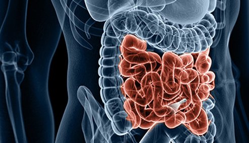
The NaviCam® SB Video Capsule Endoscopy System (AnX Robotica, Plano, TX) is the first video capsule endoscopy system that uses a deep learning model-trained AI platform, ProScan™, to assist physicians in identifying pathology in the small bowel.
Artificial Intelligence (AI) is one of the primary capabilities behind the development of precision medicine that is broadly used today. AI is not one, but rather a collection of technologies. With AI, a machine or software needs to be trained using a technique called Machine Learning (ML). ML is a method of supervised learning using training algorithms that enables machines or software to learn how to make decisions using provided data to make accurate predictions.
The more complex forms of ML involve Deep Learning (DL), which are models with many levels of variables that predict outcomes. Just like our brain identifies patterns and classifies various types of information, machines can be taught to accomplish the same tasks using deep learning algorithms. Deep learning models have shown great potential in assisting physicians in identifying and analyzing medical images, including lesions in the small bowel. While conventional reading by a physician remains the primary method for diagnosis, deep learning models can provide valuable support and enhance the accuracy and efficiency of the process.
Here are a few key benefits of deep learning models in identifying lesions in the small bowel:
- Increased Accuracy: Deep learning models can be trained on large datasets of medical images, allowing them to learn patterns and features that may not be easily noticeable to the human eye. This advanced innovation can help improve the accuracy of lesion detection and reduce the risk of misdiagnosis. ProScan was trained with over 158,000 images from close to 2,000 patients and then validated with over 113 million images from over 5,000 patients. The per lesion analysis, the sensitivity and negative predictive value of ProScan is 99.9% and 100% respectively[i].
- Consistency: Human interpretation of medical images vary based on expertise, experience, and fatigue. Deep learning models, once trained, provide consistent and standardized analysis, ensuring a reliable and reproducible evaluation of small bowel lesions.
- Time Efficiency: Conventional reading of medical images is time-consuming, requiring physicians to manually analyze and interpret each image. Deep learning models can rapidly process large volumes of images, significantly reducing the time required for lesion identification. This method of reviewing small bowel images can expedite diagnosis and facilitate timely patient care.
- Decision Support: Deep learning models serve as valuable innovated assistant support tool for physicians in lesion diagnosis. By providing additional insights and highlighting potentially suspicious areas, these models may assist physicians with clinical decisions and improve overall diagnostic accuracy.
However, it’s important to note that deep learning models are not meant to replace physicians but rather act as complementary tools. Physicians still play a crucial role in clinical decision-making, considering the model’s output alongside other patient information, medical history, and their expertise.
In summary, deep learning models can offer physicians valuable support in identifying small bowel lesions. The ability of deep learning models to analyze large volumes of data, enhance accuracy, provide consistency, and expedite the diagnostic process offers improvement to the field of medical imaging.
[i] Gastroenterology 2019;157:1044–1054
ProScan is currently under review by FDA and not available for sale in the United States.
NaviCam® SB with EsView
All small bowel capsule systems are not created equal. Using novel aspherical lens technology, which minimizes distortion, the NaviCam® SB has an image resolution of 640*480 (CMOS Imager), Variable Frame Rate of 0.5 – 6 FPS and an extended battery life over 16 hours (1fps). But what makes the NaviCam® SB System different is ProScan™ Intelligent Reading Support and is the driving reason why AnX Robotica is not just another capsule company.
ProScan™ incorporates Deep Neural Networks (DNN), such as a Convolutional Neural Network (CNN), a deep learning algorithm that can differentiate one image from another. This deep learning model enables ProScan™ to differentiate normal mucosa from abnormal lesions, which assists the user with the study review as well as significantly improving efficiency.
The ProScan feature is not intended to detect pathology in lieu of a physician and should not be used as a substitute for reviewing the entire video.
Let's get in touch!
Would you like to receive more information or do you have any questions about this innovation?
Please do not hesitate to contact us via button below. We're more than happy to schedule a call or demo with you!