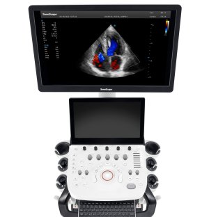
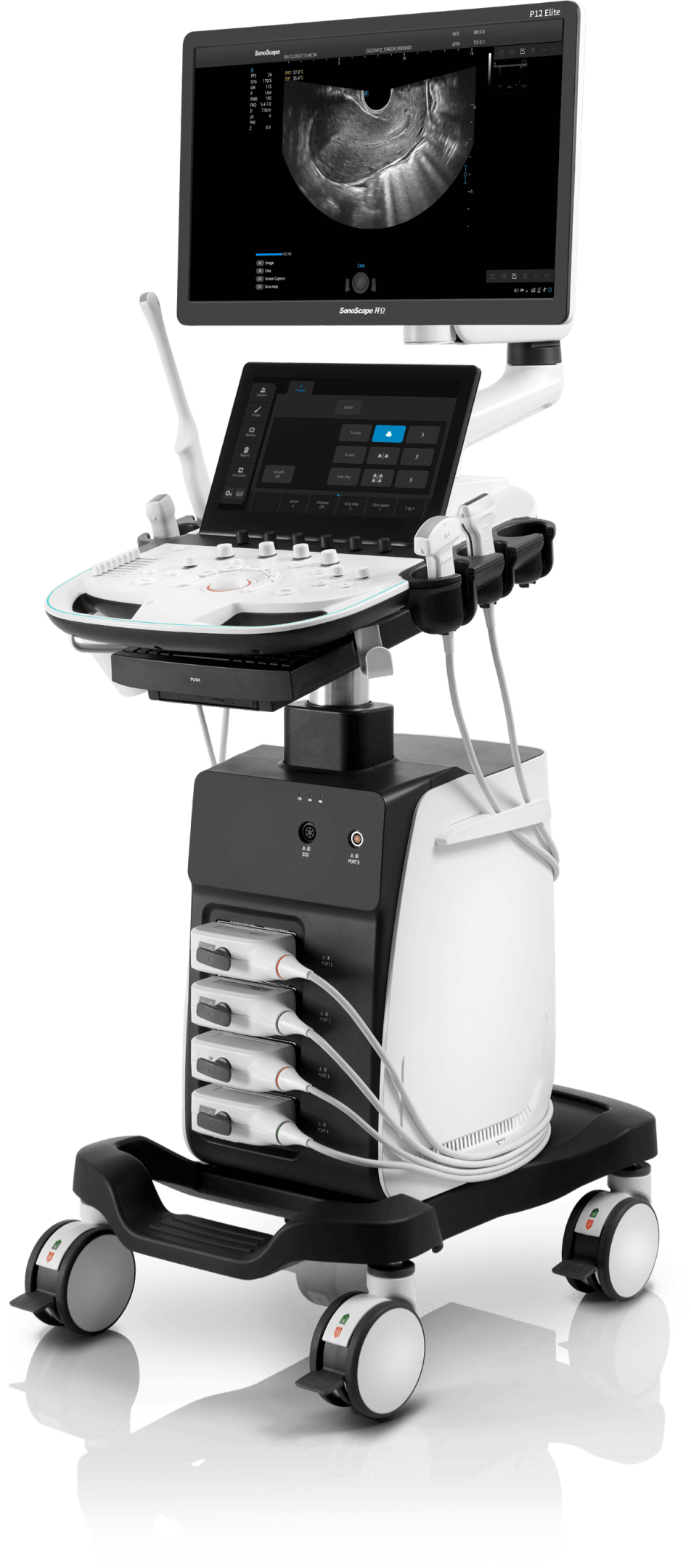
Sonoscape P12 Elite Ultrasound
To fulfill the promise of caring for life through innovation, SonoScape proudly presents the P12 Elite: a high-definition imaging solution powered by cutting-edge technologies, delivering informative details and boosting diagnostic confidence.
Description
Highlights
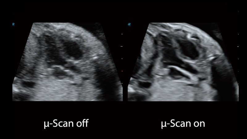
- μ-Scan+ – greatly improves visualization of organs and lesions, suppresses speckle artifacts, and maintains true tissue architecture.
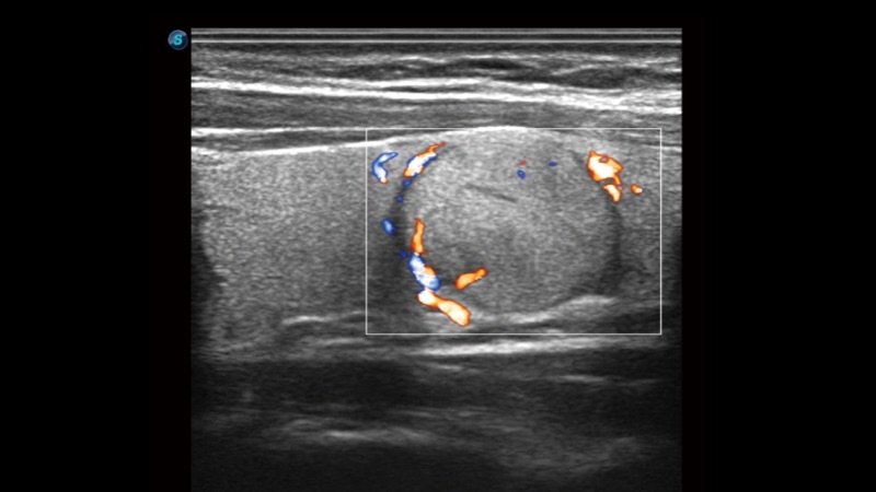
- SR Flow – enhances the detection of low-velocity flow signals, improves spatial resolution, and prevents overflow, offering accurate hemodynamic information.
- WideScan – enables real-time enlargement of ultrasound images when using linear or convex probes, providing a more comprehensive view of large lesions or structures.
- Real-time Color Panoramic – combines color flow with real-time panoramic imaging to visualize blood flow throughout an entire vein or artery. Errors can be backtracked and corrected without interrupting scanning.
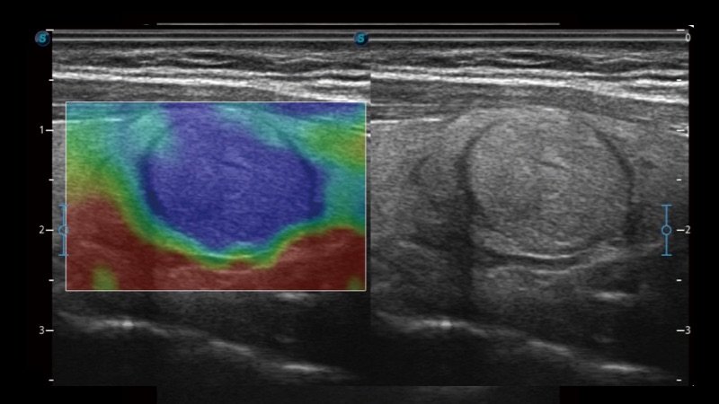
- C-xlasto Imaging – delivers quantitative elastic analysis with high reproducibility and consistency, supported by multiple probe types.
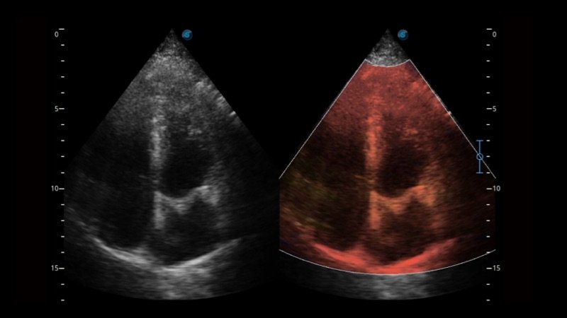
- Tissue Doppler Imaging – provides velocity and other clinical information on myocardial functions to support clinicians in analyzing and comparing motions of different cardiac regions.
- Contrast-Enhanced Ultrasound (CEUS) – offers comprehensive imaging and quantification capabilities for assessing perfusion dynamics across clinical settings. The unique Dynamic Acoustic Control technology regulates contrast agent acoustic pressure, ensuring longer duration and improved lesion perfusion in delayed phases.
- S-Fetus – based on reliable deep-learning big data algorithms, this one-click solution automatically acquires standard fetal planes and biometric results with intelligence, accuracy, and efficiency.
- Vis-Needle – enhances visualization of needle location in tissue via beam steering and deflection, minimizing surrounding tissue damage, increasing initial success rates, and reducing puncture risks.
- S-Live – delivers real-time 3D imaging of subtle anatomical structures for intuitive diagnosis and enhanced patient communication.
Interaction with Care
- Sono-Help – an inspiring tutorial system illustrating probe placement, anatomy, and sample standard images for applications across liver, kidney, cardiac, breast, thyroid, obstetrics, vascular, and more—ideal for supporting less experienced clinicians.
- Sono-Drop – enables wireless connection between P12 Elite and smartphones so ultrasound images can be shared easily with patients and their families.
- Sono-Synch – enables real-time interface and camera sharing, connecting two ultrasound systems remotely for tele-consultation and tutorials.
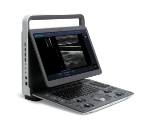
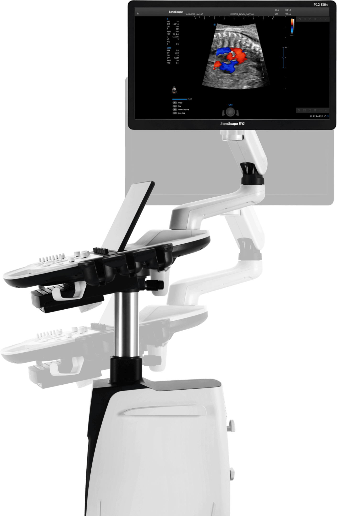
User-Friendly Design for Efficient Workflow
Related products