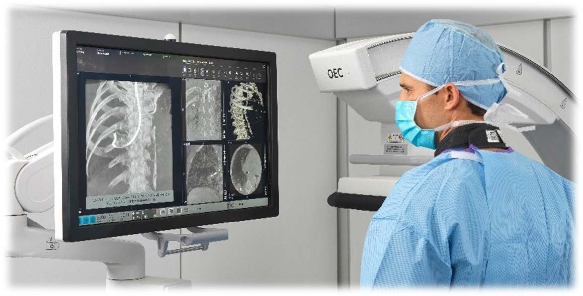
Real-time 3D imaging for bronchoscopy: OEC 3D Lung Suite
The OEC 3D Lung Suite combines 3D cone beam CT imaging with real-time fluoroscopy to make bronchoscopy procedures more accurate and efficient. Advanced visualization tools support clinicians in locating lesions and confirming instrument positioning during the procedure.
Description
Description
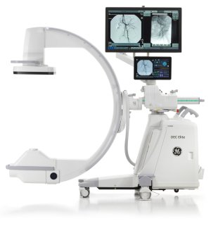
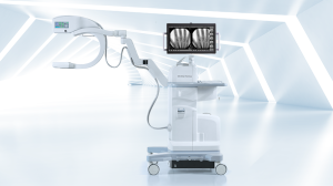
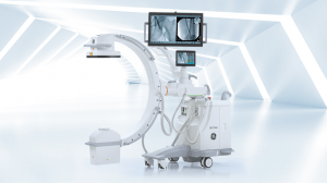
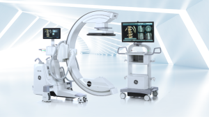
The OEC 3D Lung Suite is a software extension for the OEC 3D mobile C-arm that simplifies bronchoscopy procedures. By combining 3D imaging with live fluoroscopy, the system provides improved insight into lung anatomy and supports precise localization of target areas.
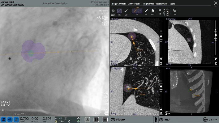
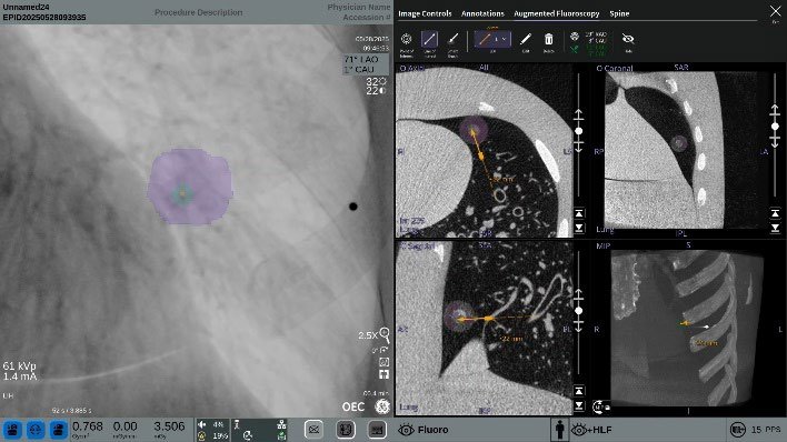
With augmented fluoroscopy, regions and reference lines from 3D images are projected directly onto live images, enabling accurate instrument positioning.
Thanks to fast 3D acquisition, flexible scan settings, and integration with navigation and robotic systems, the Lung Suite supports an efficient workflow and reduces the need for repeated imaging.
Real-time precision
Technical Services
Technical details
Imaging
- 3D cone beam CT (CBCT) imaging
- Volume size up to 19 × 19 × 19 cm
- Multiplanar visualization
Acquisition
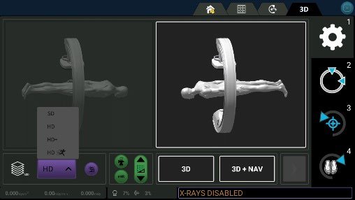
- HD Sprint mode: 16-second scan
- Up to 400 projections per scan
- Available modes: SD, HD, HD+, HD Sprint
Augmented fluoroscopy
- Overlay of 3D information on live fluoroscopy
- Target Points and Line of Interest
- Smart Brush tool
Positioning
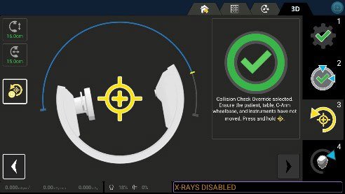
- Guidance Presets (motorized positions)
- Bullseye View (top-down view)
- Progress View (perpendicular view)
Workflow & integration
- OEC Open interface for navigation and robotics
- DICOM import (CT/MR)
- Volume Viewer with advanced analysis tools
Related products