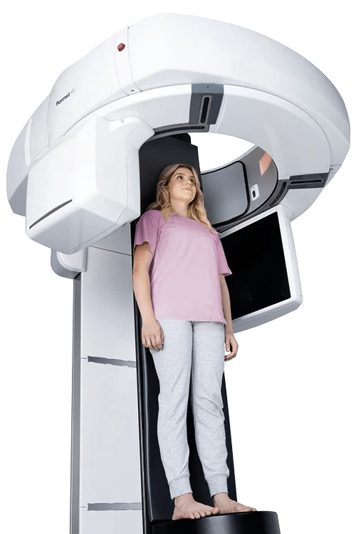
Cone-beam CT scanner – Planmed XFI®
Planmed XFI is an advanced cone-beam CT (CBCT) system that enables full 3D imaging of the entire body, from head to feet. The system supports both standing (weight-bearing) and supine scans, allowing clinicians to evaluate the musculoskeletal system in a natural, functional position.
This innovative technology provides an extensive field of view, ultra-high resolution, and low radiation doses, making it particularly well suited for orthopedic and musculoskeletal diagnostics.


Omschrijving
High-quality 3D imaging of the entire body
The Planmed XFI features a large 85 cm gantry opening and a wide flat-panel detector system. During the scan, the X-ray source and detector rotate 360° around the body, enabling detailed 3D images of the entire anatomy to be captured.
The system delivers ultra-high-resolution images down to 75 microns, providing exceptional image quality and diagnostic precision.
Key features:
- Full 3D imaging of the entire body
- Scanning in standing (weight-bearing) and supine positions
- 85 cm gantry opening for improved patient comfort
- Large field of view (FOV) for complete anatomical visualization
- Ultra-high resolution up to 75 µm
- 360° rotation for accurate volumetric imaging
Low-dose imaging with optimal image quality
In 3D imaging, it is important to combine diagnostic quality with the lowest possible radiation dose. This is particularly relevant for young patients, who are more sensitive to radiation.
The Planmed XFI supports the ALADA principle (As Low As Diagnostically Acceptable), where the radiation dose is minimized without compromising diagnostic value.
With the Planmeca Ultra Low Dose™ protocol, the effective patient dose can be reduced even further thanks to intelligent 3D reconstruction algorithms, while maintaining image quality.
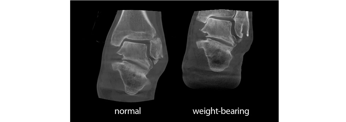
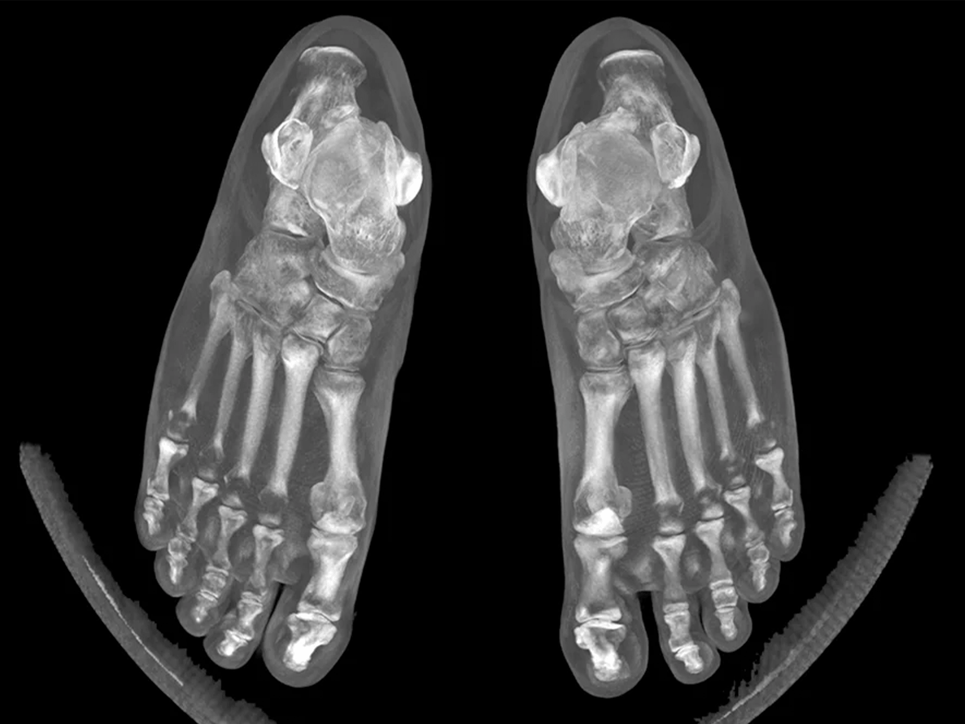
Advantages of 3D weight-bearing imaging
Applications